As you explore, examine, and evaluate the human body, musculoskeletal ultrasound offers you a window into the intricate workings of muscles, tendons, and joints. This introduction is tailored for beginners like you, who are taking their first steps into the realm of diagnostic imaging.
You’ll learn the essentials of ultrasound technology, including the basics of the equipment and the anatomy most relevant to scanning. You’ll discover how to select the right probe, position your patients correctly, and follow standard scanning protocols.
We’ll guide you through the nuances of interpreting images and identifying common pathologies. With practical advice and clear guidance, you’ll gain the confidence to use ultrasound effectively, enhancing your diagnostic acumen and paving the way for improved patient outcomes.
Welcome to the hands-on world of musculoskeletal ultrasound; let’s begin your journey.
Key Takeaways
- Musculoskeletal ultrasound relies on sound waves to generate images of soft tissue, joints, and ligaments.
- Accurate identification of key structures such as bones, tendons, and ligaments is necessary for precise imaging.
- Understanding the spatial relationships between joints and muscle tendons is essential for interpreting ultrasound scans correctly.
- Selecting the appropriate probe type is critical for accurate musculoskeletal imaging.
Everything You Need To Know About Us – Physicians Group, LLC
The expert team at Physicians Group, LLC, a specialized clinic for automobile accident injuries, offers comprehensive care across a network of 25 accessible locations.
TOUCH BASE FOR ASSISTANCE WITH YOUR APPOINTMENT ONLINE
With a widespread presence in regions including Auburndale, Bradenton, Brandon, Palmetto, Ft Myers, Jacksonville Beach, Jacksonville, Orange Park, Lakeland, Clearwater, New Port Richey, Port Charlotte, Sarasota, Sebring, Spring Hill, St. Petersburg, Tampa, Temple Terrace in Florida, as well as Brooklyn Park, Robbinsdale, Minneapolis, Richfield, and St Paul in Minnesota, Physicians Group, LLC is a prime destination for addressing injuries post-automobile accidents.
Our Expertise
Endorsed by The Joint Commission, Physicians Group, LLC stands as a comprehensive hub for the treatment of musculoskeletal injuries. Its multidisciplinary team comprises seasoned medical doctors, skilled chiropractors, osteopathic professionals, attentive nurse practitioners, and proficient physician assistants.
The group is adept at managing a spectrum of musculoskeletal discomforts, from joint, back, to neck pain. By leveraging state-of-the-art technology and diagnostic tools like DynaROM™ motion testing, digital motion X-rays (DMX) for the cervical spine, and cutting-edge regenerative therapies, they are equipped to foster optimal recovery.
Emphasizing a nurturing, patient-focused atmosphere, the dedicated personnel at Physicians Group, LLC ensures thorough consultations and in-depth discussions, allowing them to grasp every patient’s specific complaints and aspirations fully. Treatment plans emphasize non-invasive and integrative approaches, with surgical interventions being considered as necessary.
Don’t allow the aftermath of vehicle accidents to impede your lifestyle. Initiate your path to recovery at Physicians Group, LLC by reaching out to your local branch or by securing an appointment through our online platform.
Our Services:
–CHIROPRACTOR
–IMAGING
–DIAGNOSTIC TESTING
–PAIN MANAGEMENT
–PHYSICAL THERAPY
–MEDICAL EVALUATIONS
Conditions We Treat:
–CAR ACCIDENT INJURIES
–TRUCK ACCIDENT INJURIES
–MOTORCYCLE ACCIDENT INJURIES
–PEDESTRIAN ACCIDENTS
–BICYCLE ACCIDENT INJURIES
–BUS ACCIDENT INJURIES
–SLIP AND FALL INJURIES
–WORKPLACE ACCIDENT INJURIES
–RIDESHARE ACCIDENT INJURIES
Contact Us:
→ https://physiciansgroupllc.com/contact-us/
Understanding Ultrasound Basics
Comprehension of ultrasound principles is essential as you embark on mastering musculoskeletal imaging techniques. Musculoskeletal ultrasound relies on sound waves to generate images of soft tissue, joints, and ligaments.
You’ll need to understand frequency and wavelength, as higher frequencies produce finer resolution but have reduced penetration. It’s a balance; you must choose the appropriate transducer for the specific body part and condition you’re assessing.
You’ll also manipulate the gain to optimize image quality. Gain increases signal strength, but excessive gain can lead to image distortion, so you must adjust it carefully.
Be methodical in your approach to each scan, moving the transducer slowly to maintain image clarity. Remember, precision in these foundational concepts is paramount for accurate diagnosis and successful treatment planning.
Anatomy Essentials for Scanning
Before you initiate scanning, it’s critical that you’re familiar with the anatomical landmarks.
You’ll need to accurately identify key structures such as bones, tendons, and ligaments to ensure precise imaging.
Understanding the spatial relationships between joints and muscle tendons is essential for interpreting ultrasound scans correctly.
Identifying Key Structures
To effectively navigate musculoskeletal ultrasound, you’ll need to identify several critical anatomical landmarks. Begin by pinpointing bones; they serve as primary reference points due to their high echogenicity, which makes them appear bright on the ultrasound screen.
Next, differentiate between tendons and ligaments—both are hyperechoic structures, but tendons often change shape as they move, while ligaments remain static.
Pay close attention to muscle tissue, recognizing its fibrillar pattern and lower echogenicity compared to tendons and ligaments.
Lastly, don’t overlook the importance of identifying bursae and nerves; bursae appear as anechoic or hypoechoic spaces, whereas nerves present a honeycomb-like structure.
Your precision in recognizing these structures is paramount for accurate diagnosis and effective treatment planning.
Joint Space Visualization
Once you’ve become adept at identifying key anatomical landmarks, you’ll want to turn your attention to the subtleties of joint space visualization, a critical skill in musculoskeletal ultrasound. This involves discerning the narrow gaps between adjacent bones, which constitute the joint spaces. You must systematically analyze these areas for signs of pathology, such as effusions or synovial thickening.
Start with a clear understanding of the normal joint anatomy. Recognize that joint spaces may vary widely in size and shape among different joints and individuals. Use a high-frequency linear transducer for superior resolution. Adjust the ultrasound machine settings to optimize the contrast between the hypoechoic (dark) joint fluid and the hyperechoic (bright) bone surfaces.
Careful manipulation of the transducer will help you avoid anisotropy, ensuring accurate visualization of joint spaces.
Muscle Tendon Relationships
Understanding muscle tendon relationships is crucial, as you’ll often be assessing these structures for signs of tears, tendinopathy, or other abnormalities. Muscles and tendons have a symbiotic relationship where the muscle transitions into a tendon, attaching to bone to enable movement. You must be methodical in identifying the echotexture of muscles, which typically appears as a feathery pattern, juxtaposed with the fibrillar pattern of tendons on ultrasound imaging.
Here’s a table to illustrate the key differences:
| Structure | Appearance on Ultrasound | Common Pathologies |
| Muscle | Feathery pattern | Strain, Tear |
| Tendon | Fibrillar pattern | Tendinopathy, Tear |
| Bone | Hyperechoic with shadow | Fracture, Spur |
Analyzing these relationships with precision will enhance your scanning technique and diagnostic accuracy.
Equipment and Probe Selection
You’ll find that selecting the appropriate probe type is critical for accurate musculoskeletal imaging.
Assessing image quality factors ensures you capture the clearest possible representations of tissues.
Lastly, adhering to strict equipment maintenance practices is paramount for the longevity and reliability of your ultrasound system.
Probe Types
When selecting a probe for musculoskeletal ultrasound, it’s crucial to consider the type of tissue and depth you’ll be examining. Your choice will directly influence the quality of the images and, consequently, the accuracy of the diagnosis. Various probes are designed to optimize the visualization of specific anatomical structures:
- Linear probes: Ideal for high-frequency imaging of superficial structures, such as tendons and ligaments.
- Curved array probes: Better suited for deeper tissues due to their lower-frequency capabilities, useful for larger patients.
- Phased array probes: Typically used in cardiac imaging but may provide a small footprint for navigating challenging areas.
- Hockey stick probes: Small and highly maneuverable, perfect for intricate areas like fingers and toes.
Each probe has unique attributes that can enhance your diagnostic precision when matched with the appropriate application.
Image Quality Factors
Selecting the right probe and equipment is crucial for enhancing the clarity and detail of musculoskeletal ultrasound images. This choice directly affects the resolution and depth of field, which are essential for discerning fine structures within the musculoskeletal system.
For superficial tissues, a high-frequency linear probe is recommended as it yields finer detail. On the other hand, a lower-frequency probe is better for visualizing deeper structures as it can penetrate more effectively.
In addition to choosing the appropriate probe, it is important to ensure that your ultrasound machine is calibrated and has sufficient processing power to handle the data-intensive demands of high-resolution imaging.
Adjusting the gain thoughtfully is also important. Setting it too high can create noise, which obscures detail, while setting it too low may cause subtle findings to be missed.
Another important aspect of image acquisition is methodically fine-tuning the focus to the region of interest. This directly sharpens the image and helps in capturing the desired structure with clarity.
Equipment Maintenance Practices
While selecting the appropriate probe is crucial for high-quality images, maintaining your equipment ensures its longevity and consistent performance. You’re tasked with safeguarding your investment by adhering to a strict maintenance schedule. This involves meticulous cleaning, proper storage, and routine checks for wear and tear.
To delve deeper, consider the following practices:
- Regularly disinfect probes using approved solutions to prevent cross-contamination.
- Inspect the transducer’s lens for micro-abrasions that can degrade image quality.
- Calibrate your system periodically to ensure accuracy in diagnostics.
- Store equipment in a dust-free environment to protect sensitive components.
Embrace these protocols to optimize the functionality of your ultrasound equipment. Your analytical approach to maintenance will pay dividends in the reliability and precision of your musculoskeletal imaging procedures.
Patient Positioning Techniques
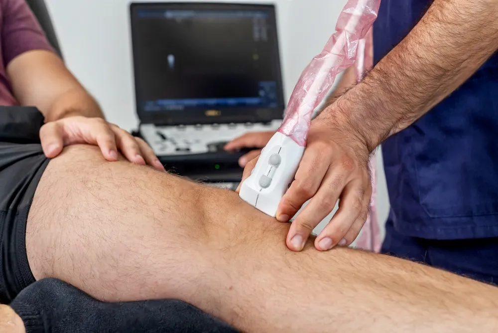
You’ll need to position your patient properly to ensure the ultrasound probe has optimal access to the area of interest. Begin by analyzing the target structure’s anatomical location. If it’s superficial, ask the patient to adopt a position that brings the area closer to the surface. For deeper structures, choose a position that reduces intervening tissue.
Ensure the patient is comfortable and stable to minimize movement artifacts. Use pillows or foam wedges for support if necessary. Remember, the ultimate goal is a clear image; this often means adjusting the patient’s position minutely until the desired view is achieved. Be methodical in these adjustments, always observing the impact on the ultrasound image, and make precise corrections to enhance visualization.
Standard Scanning Protocols
Understanding standard scanning protocols is crucial as they provide a systematic approach to capturing comprehensive musculoskeletal images. These protocols help ensure that you don’t miss any critical structures and maintain consistency across examinations.
Here are key elements to consider:
- Transducer Selection: Choose the appropriate transducer based on the depth and resolution required for specific tissues.
- Image Orientation: Consistently orient the images with anatomical landmarks to facilitate accurate interpretation.
- Scanning Sequence: Follow a predetermined sequence to assess all relevant anatomical structures systematically.
- Documentation: Record images and measurements precisely, noting any deviations from normal findings.
Interpreting Ultrasound Images
Once you’ve captured your musculoskeletal ultrasound images, it’s essential to accurately interpret the various shades of gray representing different tissues. Your analytical skills are crucial here as you discern subtle differences. Bone surfaces will appear hyperechoic, with a bright white surface, while tendons and nerves present as fibrillar structures with a less bright, but still white, echogenicity. Muscles and soft tissues show as shades of gray, and fluid collections, as anechoic or black areas.
Here’s a table to help you systematically approach the images:
| Tissue Type | Echogenicity | Characteristic Appearance |
| Bone | Hyperechoic | Bright white, linear |
| Cartilage | Hypoechoic | Homogeneous gray |
| Muscle | Intermediate | Speckled gray pattern |
| Tendon | Hyperechoic | Fibrillar, less bright |
| Fluid | Anechoic | Black, no internal echoes |
Examine these echogenic patterns carefully and compare them to adjacent structures for an accurate diagnosis.

Common Pathologies Identified
Identify common musculoskeletal pathologies such as tendonitis, ligament sprains, and muscle tears by recognizing their distinct ultrasound signatures.
When you’re interpreting ultrasound images, look for specific indicators that signal these conditions:
- Tendonitis: You’ll often see a swollen and hypoechoic (darker) appearance of the tendon, sometimes accompanied by calcifications.
- Ligament Sprains: These are characterized by a discontinuity in the fibers or an abnormal ligamentous structure, with potential fluid accumulation.
- Muscle Tears: Look for a disruption in the normal muscle fiber pattern and the presence of a hematoma.
- Joint Effusions: Excess fluid within a joint capsule is usually evident as an anechoic or hypoechoic space.
Each pathology exhibits a unique pattern, making a methodical approach to image analysis crucial.
Tips for Improved Accuracy
Mastering the techniques of musculoskeletal ultrasound can significantly enhance your diagnostic precision, ensuring you’re accurately interpreting the subtle cues of various pathologies. To improve accuracy, you must maintain a methodical approach.
Begin by selecting the appropriate transducer and frequency for the anatomical area of interest; high-frequency linear transducers are typically ideal for superficial structures. Ensure optimal patient positioning to expose the target area and minimize patient discomfort, which can affect image quality.
Adjust the gain settings meticulously to avoid over or under-saturation, which can obscure critical details. Use a systematic scanning technique, sweeping the transducer slowly to examine the structure in multiple planes and angles. This reduces the risk of missing lesions or abnormalities.
Consistently apply a light but firm pressure to maintain contact without compressing tissues, as excessive force can distort the architecture you’re striving to assess.
Frequently Asked Questions
How Does Musculoskeletal Ultrasound Compare to MRI in Terms of Diagnostic Accuracy for Various Injuries?
You’re considering musculoskeletal ultrasound versus MRI for injury diagnosis. Generally, MRI offers greater detail, but ultrasound provides real-time imaging and is more cost-effective, though its accuracy can vary based on the operator’s skill.
Can Musculoskeletal Ultrasound Be Used for Guiding Therapeutic Procedures, and if So, Which Ones?
Yes, you can use musculoskeletal ultrasound to guide therapeutic procedures, such as injections for joint pain, tendon repairs, and aspiration of fluid collections. It provides real-time imaging, improving accuracy and safety.
What Are the Limitations of Musculoskeletal Ultrasound in Obese Patients or Those With Significant Muscle Mass?
In obese patients or those with significant muscle mass, you’ll find musculoskeletal ultrasound less effective due to poor sound wave penetration, resulting in suboptimal images and potentially less accurate diagnoses.
How Can One Maintain and Ensure the Hygiene of the Ultrasound Equipment, Especially in a Busy Clinic Setting?
You’ll need to regularly disinfect the ultrasound probes between patients and use disposable covers. Implement a strict cleaning schedule and ensure staff are trained in proper sanitation protocols to maintain equipment hygiene.
What Kind of Training and Certification Is Required for a Healthcare Professional to Perform Musculoskeletal Ultrasounds?
You’ll need specific training in musculoskeletal sonography and certification from a recognized body, like the ARDMS, to perform musculoskeletal ultrasounds competently and legally within your healthcare practice.
Conclusion
In conclusion, you’ve now navigated the fundamentals of musculoskeletal ultrasound. From mastering anatomy to selecting the right equipment and adopting proper scanning protocols, you’re equipped to discern common pathologies with greater precision.
Remember, consistent practice hones your interpretive skills. So, keep refining your technique, stay methodical in your approach, and you’ll see your accuracy improve.
Your journey into musculoskeletal imaging is just beginning, and your dedication will be the key to your success.